Common fundus disorder:
-
CNV - Choroidal neovascularization
-
DR - Diabetic Retinopathy
-
AMD - Age-related macular degeneration
-
PCV - Polypoid Choroidal Vasculopathy
-
CSC - Central Serous Retinopathy
-
Retinal Cleft Palate
-
Macular Hole
-
Vitreous Macular Traction
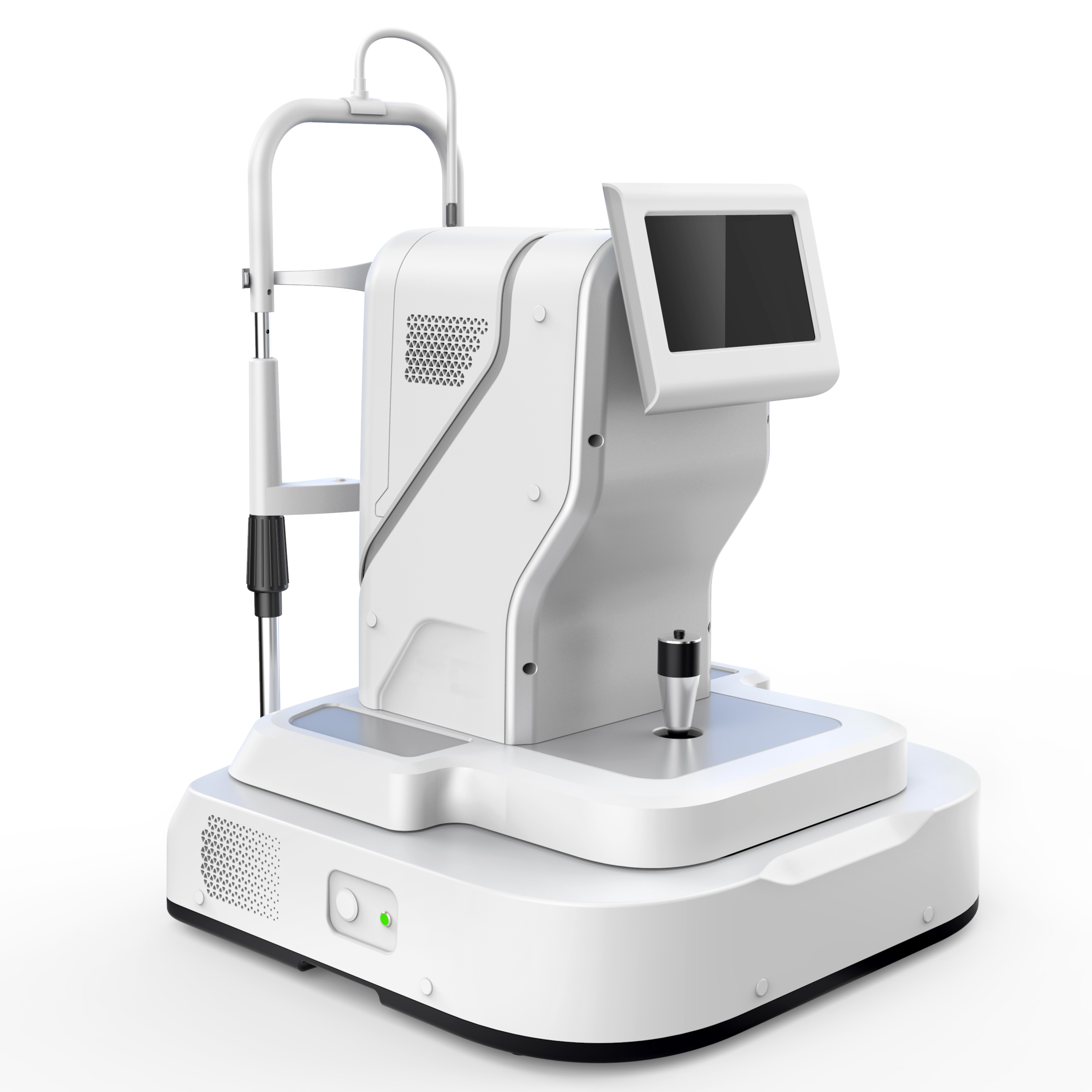
Diagnosis of Fundus Disease
It accurately identifies retinal disorders, helps to screen and reduce the missed diagnosis of retinal diseases at the outpatient clinic, and can greatly improve the efficiency of clinical use.
Optimized functions, popular and practical
-
Flexible optics parts, easy to operate, easy access to patient fundus images
-
Easy to use, intuitive, fast focus
-
One-click collection saves outpatient time
Smart Reading (Optional)
A fundus disease intelligence software developed based on deep learning to indentify specific structures of retinal images faster and more efficiently.
| Measurement |
Axial resolution:6μm (in tissue)
Horizontal resolution: 13μm
|
| Scanning |
Maximum A scanning speed: 20KHz, tolerance ± 5%
Maximum scanning depth: 2.65mm (tissue), tolerance ± 3%
Maximum scaning range: 13mm x 13mm, tolerance ± 5%
|
| Light Source |
Center wavelength: 840nm, tolerance ± 10nm
Half width: 35nm, tolerance ±5nm
Optical Power: ≤ 750 μw (at the cornea)
Refractive compensation range: ≥ -12D ~ +12D
|
| Fundus Image Light Source |
Center wavelength 760nm, tolerance ± 5nm
Optical power: ≤50μw
|
| Retinal Thickness Measurement |
Retinal thickness measurement tolerance: ± 3% (in tissue)
Retinal thickness measurement repeatability: ≤2%
|